

A-Z™ Autism Drug Discovery Platform
Creative Biolabs provides the A-ZTM Autism Drug Discovery Platform for research programs that need to connect an ASD-relevant hypothesis with a qualified model, a measurable phenotype, candidate screening, and translational validation.
 Fig.1 A-Z™ Autism Drug Discovery Platform.
Define the Discovery Question and Success Criteria
ASD is biologically heterogeneous, so the same model and endpoint should not be applied to every program. A target-validation study centered on a high-confidence risk gene requires a different evidence chain from a phenotypic screen in idiopathic patient-derived neurons. Likewise, a compound intended to normalize network excitability needs functional measurements that are not interchangeable with a viability or marker-only assay. We begin by translating the therapeutic hypothesis into a testable sequence of decisions, controls, and advancement criteria.
Typical entry points include:
- Target and pathway validation: Determine whether genetic correction, knockdown, knockout, overexpression, or pharmacological modulation changes a reproducible ASD-associated cellular or molecular phenotype.
- Model development: Generate or qualify patient-derived, mutation-specific, isogenic, cellular, organoid, or animal models that preserve the biology needed for the intended experiment.
- Phenotypic hit discovery: Identify compounds that normalize a defined feature of neurogenesis, morphology, synapse formation, calcium signaling, neuronal firing, network organization, glial response, or another project-specific phenotype.
- Candidate ranking: Confirm concentration-dependent activity, separate efficacy from cytotoxicity or assay interference, assess selectivity, and compare performance across genotypes, donors, cell batches, or model tiers.
- Mechanism and biomarkers: Connect an observed rescue to target engagement, pathway modulation, molecular signatures, or functional biomarkers that can support later studies.
- Translational de-risking: Integrate exposure, pharmacodynamics, tolerability, and efficacy in a fit-for-purpose in vivo or ex vivo context when a program is ready to move beyond discovery-stage human cell models.
A program may begin with the broader Autism Disorder Drug Discovery Service or move directly into Autism Spectrum Disorder Genetic Model Development when the genotype and desired model are already defined. The study plan identifies the primary decision endpoint, supporting readouts, biological and technical replicates, acceptance ranges, and the conditions under which a result will be advanced, repeated, or stopped.
Build an ASD Model Portfolio That Matches the Biology
Human iPSC-derived systems can retain donor genetics and enable isogenic correction or mutation introduction, while 2D cultures offer scale and direct access to individual cell types. Three-dimensional systems add developmental organization and multicellular interactions but require tighter control of batch, size, composition, and maturation. In vivo studies can address integrated exposure, circuit, and behavioral outcomes, yet their relevance depends on the construct and phenotype being modeled.
| Model tier | Best suited for | Representative endpoints | Qualification priorities |
| Patient-derived or engineered iPSC models | Genotype-specific mechanisms, isogenic comparisons, target validation, personalized-response studies | Neurogenesis, lineage balance, neurite morphology, synaptic markers, pathway activity | Identity, pluripotency, genomic integrity, edit confirmation, differentiation consistency |
| 2D neural cultures and co-cultures | Scalable phenotypic screens, cell-autonomous and glial-interaction studies, concentration-response testing | Viability, proliferation, calcium dynamics, synaptogenesis, cytokines, high-content morphology | Cell composition, maturity, assay window, plate effects, positive and negative controls |
| Neural spheroids, cortical organoids, and assembloids | Developmental organization, cell-cell interactions, migration, circuit formation, longer-term exposure | Layer or lineage markers, spatial phenotypes, network activity, transcriptomic signatures | Size and morphology, batch reproducibility, regional identity, necrosis, functional maturation |
| Target-based and reporter systems | Focused mechanism testing, pathway modulation, binding or enzymatic hypotheses | IC50 or EC50, target engagement, reporter response, selectivity, pathway rescue | Construct validation, dynamic range, interference controls, orthogonal confirmation |
| In vivo ASD models | Exposure-response relationships, behavioral or circuit-level confirmation, translational de-risking | Project-defined behavior, pharmacodynamic biomarkers, tissue exposure, tolerability | Genotype or induction verification, baseline phenotype, randomization, blinding, cohort power |
Human Cellular and Three-Dimensional Models
Human cellular programs may use existing donor lines, newly generated lines, or engineered isogenic pairs. Our iPSC-Derived Neuron and NPC Generation capabilities can support cortical or other project-defined neural differentiation, while iPSC Characterization can document identity, pluripotency, genomic status, and other agreed quality attributes. Depending on the hypothesis, studies may compare excitatory and inhibitory neurons, neural progenitors, astrocytes, microglia, or defined multicellular systems. The Neuron, Astrocyte and Microglia Co-Culture Assay is relevant when a phenotype may depend on glial signaling, inflammatory tone, trophic support, or cell-cell feedback rather than neuronal genotype alone.
For spatial and developmental phenotypes, the Brain Organoid Modeling Service can be integrated with histology, imaging, transcriptomics, and functional recording. Organoids or assembloids may be selected when the project needs to examine progenitor behavior, lineage allocation, neuronal migration, cortical organization, excitatory-inhibitory balance, synaptic maturation, or genotype-dependent network development. Model complexity is increased only when the additional biological context can change the project decision.
Genetic and In Vivo Models
Genetic strategies may include knockout, knock-in, conditional, point-mutation, copy-number, rescue, or reporter designs, subject to project feasibility. Isogenic comparisons reduce background variation and are especially useful for causality and target-engagement studies; donor panels are valuable for testing penetrance, heterogeneity, and response stratification. When integrated physiology or behavioral confirmation is required, the Autism Spectrum Disorder Mouse Model Development Service can support a project-defined model and phenotyping strategy.
Qualify Assay-Ready Phenotypes Before Screening
A screen is informative only when the starting phenotype is stable, measurable, and relevant to the therapeutic hypothesis. Qualification therefore precedes scale-up. We compare disease and control conditions across independent differentiations, cell preparations, organoid batches, plates, or cohorts; test whether the phenotype persists within the operational assay window; and determine whether known or mechanistically relevant controls move the endpoint in the expected direction.
Representative qualification domains include:
- Model identity and state: Cell-line authentication, genotype confirmation, pluripotency or lineage markers, cell composition, differentiation stage, organoid regional identity, and culture health.
- Developmental phenotypes: Progenitor proliferation, cell-cycle exit, lineage allocation, neurogenesis, migration, neurite extension, dendritic complexity, and maturation kinetics.
- Synaptic and network function: Synaptic density, excitatory and inhibitory markers, spontaneous firing, bursting, synchrony, network connectivity, stimulus response, and short-term plasticity.
- Cellular signaling: Calcium dynamics, cAMP or pathway reporters, receptor or ion-channel function, phosphorylation, transcriptional response, and target-engagement markers.
- Glial and neuroimmune biology: Astrocyte support, microglial activation or phagocytosis, cytokine or chemokine release, complement-related signals, and effects of co-culture composition.
- Assay performance: Signal window, variability, edge and plate effects, Z-prime or another agreed robustness statistic, control separation, reproducibility, throughput, and sample integrity.
Endpoint development can draw on Neurogenesis Assays, Synaptogenesis Assays, Neurite Outgrowth and Degeneration Assays, and Cell Viability Assays. Primary and counterscreen endpoints are deliberately separated: for example, a morphology rescue may be paired with cell count, membrane integrity, or apoptosis so an apparent improvement is not driven by selective loss of a subpopulation.
Screen Candidates and Resolve Mechanism
A primary assay may test a focused set of pathway modulators, a repurposing collection, a broader small-molecule library, biologics, peptides, oligonucleotides, or genetic perturbations. Hits are then retested from independent source material, evaluated across a concentration range, and confirmed with an orthogonal method. Counter-screens address cytotoxicity, nonspecific fluorescence, reporter interference, generalized suppression of activity, and effects in matched non-disease cells.
Phenotypic and Functional Screening
The Neuronal High-Content Imaging Assay can quantify multiparametric morphology, marker localization, cell composition, synaptic features, and survival at single-cell resolution. Calcium Imaging Assays can measure stimulus-dependent or spontaneous calcium dynamics, while the Neuronal MEA Assay can capture firing rate, bursting, synchrony, network organization, and treatment-dependent changes over time. Assays may be run as single-endpoint screens or as an integrated phenotypic signature when no single measurement adequately represents the disease state.
A typical screening sequence includes assay miniaturization and plate-map design; pilot evaluation of controls and concentration ranges; primary screening with predefined QC rules; confirmation using fresh material; concentration-response analysis; counterscreening; orthogonal validation; and cross-model testing in additional donors, isogenic pairs, 3D systems, or relevant cell types. For combination programs, single-agent ranges are established first, followed by a matrix, fixed-ratio, or sequence design and a prespecified additivity or synergy model.
Mechanism, Selectivity, and Biomarker Development
Follow-up can evaluate direct target engagement where feasible, pathway activity, genetic epistasis or rescue, temporal ordering, subcellular localization, protein or transcript changes, secreted factors, and correlation between molecular and functional responses. Comparing sensitive and insensitive genotypes or donors can reveal response modifiers and nominate biomarkers for stratification. When a candidate changes network activity, the program also determines whether the shift represents normalization, sedation-like suppression, generalized excitation, altered viability, or a durable correction of the disease-associated pattern.
Programs focused on candidate rescue can also incorporate Neuroprotective Agent Screening where the relevant ASD model includes a quantifiable vulnerability or stress-response component. All claims remain limited to the model, endpoint, and tested conditions; observations from one genotype or developmental stage are not generalized across the ASD spectrum without supporting evidence.
Related Research
Cortical Organoids Reveal Developmental Convergence across Genetically Defined ASD Forms
Gordon and colleagues assembled a large human iPSC collection representing eight ASD-associated mutations, idiopathic ASD, and unaffected controls. After quality control, 70 lines from 55 individuals were used to generate human cortical organoids and profile RNA expression at four developmental time points. Mutation-specific effects were strongest early, whereas shared transcriptional dysregulation increased as organoids matured. The study also used CRISPR interference to test candidate regulators.
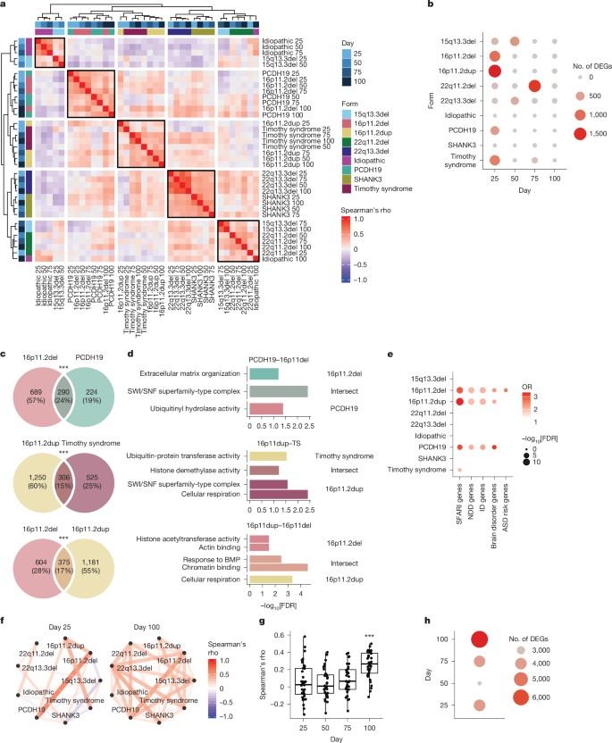 Fig. 2 Clustering and overlap of gene-expression changes across ASD cortical organoid models and developmental stages.1,3
Patient-Derived Brain Organoids Provide Subtype-Specific Functional Signatures
Perets and colleagues compared brain organoids derived from 11 individuals with ASD, including five monogenic subtypes and one idiopathic case, with organoids from four neurotypical control individuals. The investigators quantified 18 electrophysiological features and found subtype-specific differences in resting activity, evoked responses, plasticity, and network dynamics. Their resting-state data showed that ASD-derived organoids differed from controls in at least one core electrophysiological parameter, but the direction and magnitude varied across subgroups. This evidence argues for multiparametric MEA signatures, within-subtype replication, and functional stratification when evaluating candidate rescue rather than treating ASD as a single electrophysiological phenotype.
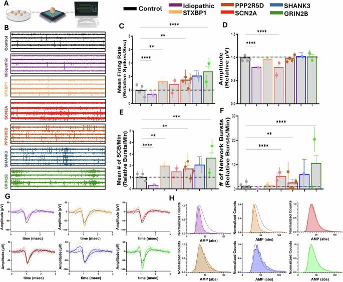 Fig. 3 Resting-state electrophysiological features of control and ASD patient-derived brain organoids.2,3
Frequently Asked Questions
- What is included in the A-Z™ Autism Drug Discovery Platform?
- Which autism spectrum disorder models can be used?
- Can patient-specific and isogenic ASD models be compared?
- How is an ASD phenotype qualified before screening?
- Which readouts are available for autism drug screening?
- Can the platform support high-content and electrophysiological screening?
- Can in vitro findings be advanced to in vivo validation?
- What information is needed to plan an autism drug discovery project?
References
- Gordon, Aaron, et al. "Developmental Convergence and Divergence in Human Stem Cell Models of Autism." Nature, vol. 651, 2026, pp. 707–719. https://doi.org/10.1038/s41586-025-10047-5
- Perets, Nisim, et al. "Patient-Derived Brain Organoids Reveal Divergent Neuronal Activity across Subpopulations of Autism Spectrum Disorder." Translational Psychiatry, vol. 16, 2026, article 164. https://doi.org/10.1038/s41398-026-03890-1
- Distributed under Open Access license CC BY 4.0, without modification.
Case Studies
- iNeuMab™ Mouse Anti-SHANK3 Monoclonal Antibody (CBP929) (Cat#: NAB-0720-Z3477)
- iNeuMab™ Mouse Anti-EFNB2 Monoclonal Antibody (CBP1159) (Cat#: NAB-0720-Z4396)
- Mouse Anti-SCN5A Monoclonal Antibody (CBP708) (Cat#: NAB-0720-Z2720)
- iNeuMab™ Rabbit Anti-Alpha-synuclein (CBP1631) (Cat#: NAB-08-PZ079)
- iNeuMab™ Mouse Anti-LRP1 Monoclonal Antibody (CBP3363) (Cat#: NAB-0720-Z6479)
- iNeuMab™ Anti-F-Spondin/SPON1 Antibody, Clone 3F4 (Cat#: NRZP-0822-ZP4740)
- iNeuMab™ Rabbit Anti-LRRK2 Monoclonal Antibody (CBP1887) (Cat#: NAB-08-PZ735)
- Mouse Anti-Human α-Synuclein Phospho (Tyr39) (CBP3706) (Cat#: NAB201250LS)
- Mouse Microglia Cell Line BV-2, Immortalized (Cat#: NCL2110P153)
- Human Glial (Oligodendrocytic) Hybrid Cell Line (MO3.13) (Cat#: NCL-2108P34)
- Mouse Midbrain Dopaminergic Neuron Cell MN9D (Cat#: NCL2110P059)
- Human Astrocytes (Cat#: NCC20-9PZ01)
- Human Dental Pulp Stem Cells (Cat#: NRZP-1122-ZP113)
- Mouse Glioma Cell Line GL261 (Cat#: NCL-2108P28)
- Human Blood Brain Barrier Model (Cat#: NCL-2103-P187)
- Green Fluorescent Tau cell Line (Cat#: NCL2110P219)
- iNeu™ Human Oligodendrocyte Progenitor Cells (OPCs) (Cat#: NCL-2103-P49)
- Human Brain Microvascular Endothelial Cells (Cat#: NCL-2103-P133)
- Alpha Synuclein Aggregation Kit (Cat#: NRZP-1122-ZP15)
- Amyloid beta 1-42 Kit (Cat#: NRP-0322-P2170)
- Human Tau Aggregation Kit (Cat#: NRP-0322-P2173)
- Human GFAP ELISA Kit [Colorimetric] (Cat#: NPP2011ZP383)
- Human Poly ADP ribose polymerase,PARP Assay Kit (Cat#: NRZP-1122-ZP62)
- Beta Amyloid (1-42), Aggregation Kit (Cat#: NRZP-0323-ZP200)
- Alpha-Synuclein Aggregation Assay Kit (Cat#: NRZP-1122-ZP37)
- Beta Amyloid (1-40), Aggregation Kit (Cat#: NRZP-0323-ZP199)
- VSV-eGFP (Cat#: NTA-2011-ZP20)
- Dextran, NHS Activated (Cat#: NRZP-0722-ZP124)
- AAV2 Full Capsids, Reference Standards (Cat#: NTC2101070CR)
- ABCA1 Antisense Oligonucleotide (NV-2106-P27) (Cat#: NV-2106-P27)
- Lenti of Human TAR DNA binding protein (TARDBP) (NM_007375) ORF clone, mGFP Tagged (Cat#: NEP-0521-R0832)
- Human superoxide dismutase 3, extracellular (SOD3) (NM_003102) ORF clone, Untagged (Cat#: NEP-0521-R0808)
- Mouse SOD1 shRNA Silencing Adenovirus (Cat#: NV-2106-P14)
- Mouse Parkinson disease (autosomal recessive, early onset) 7 (Park7) (NM_020569) clone, Untagged (Cat#: NEP-0621-R0133)
- Human presenilin 1 (PSEN1), transcript variant 2 (NM_007318) ORF clone, TurboGFP Tagged (Cat#: NEP-0421-R0140)
- Human huntingtin-associated protein 1 (HAP1) transcript variant 2 (NM_177977) ORF clone, Myc-DDK Tagged (Cat#: NEP-0521-R0676)
- Human apolipoprotein E (APOE) (NM_000041) ORF clone, Untagged (Cat#: NEP-0421-R0232)
- App Rat amyloid beta (A4) precursor protein (App)(NM_019288) ORF clone, Untagged (Cat#: NEP-0421-R0053)
- Tau Antisense Oligonucleotide (Cat#: NV-2106-P29)
- NeuroBiologics™ Human Cerebrospinal Fluid (Cat#: NRZP-0822-ZP491)
- NeuroBiologics™ Monkey Cerebrospinal Fluid (Cat#: NRZP-0822-ZP495)
- NeuroBiologics™ Pig Cerebrospinal Fluid (Cat#: NRZP-0822-ZP498)
- NeuroBiologics™ Mouse Cerebrospinal Fluid (Cat#: NRZP-0822-ZP497)
- NeuroBiologics™ Rat Cerebrospinal Fluid (Cat#: NRZP-0822-ZP496)
- NeuroPro™ Anti-IDUA BBB Shuttle Protein (Cat#: NRZP-0423-ZP502)
- NeuroPro™ Anti-GDNF BBB Shuttle Protein (Cat#: NRZP-0423-ZP500)
- NeuroPro™ Anti-SGSH BBB Shuttle Protein (Cat#: NRZP-0423-ZP505)
- NeuroPro™ Anti-GDNF BBB Shuttle Protein (Cat#: NRZP-0423-ZP509)
- NeuroPro™ Anti-EPO BBB Shuttle Protein (Cat#: NRZP-0423-ZP508)
- NeuroPro™ Anti-idursulfase BBB Shuttle Protein (Cat#: NRZP-0423-ZP497)
- NeuroPro™ Anti-Erythropoietin BBB Shuttle Protein (Cat#: NRZP-0423-ZP499)
- NeuroPro™ Anti-TNFR BBB Shuttle Protein (Cat#: NRZP-0423-ZP510)
- NeuroPro™ Anti-IDS BBB Shuttle Protein (Cat#: NRZP-0423-ZP503)
- NeuroPro™ Anti-IDUA BBB Shuttle Protein (Cat#: NRZP-0423-ZP498)

